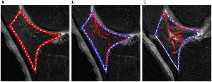
Fig. 1.
Segmentation and measurements of IPFP signal intensity alteration using MATLAB.
The segmentation and measurements of infrapatellar fat pad (IPFP) signal intensity alteration based on sagittal planes of fat-saturated T2-weighted images using MATLAB (The MathWorks, Natick, MA, USA). (A) The IPFP was segmented semi-automatically first. An initial lasso consisting of a set of points was manually created around the outer contour of IPFP and then automatically contracted inward to approximate the actual outline of IPFP. (B) The high signal intensity regions (red areas) were obtained automatically after outlining the boundary of IPFP. (C) The clustering effect of high signal intensity was greater than that in (B).

