
Figure 5.
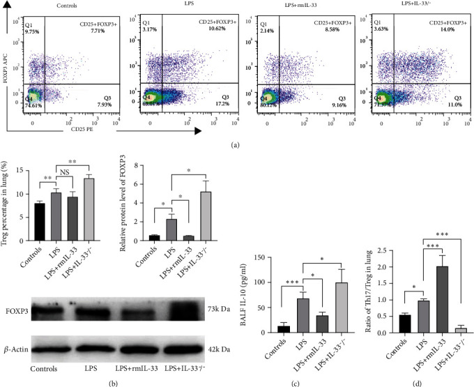
IL-33 deficiency enhanced the Treg cell response in murine ARDS. (a) The percentage of FOXP3+CD25+CD4+ T cells in the lung was detected by flow cytometry. (b) FOXP3 protein expression in lung tissue was measured by Western blotting. (c) The concentrations of IL-10 in BALF were detected by ELISA. (d) The ratio of Th17/Treg cells in the lung. The data are expressed as the means ± SDs. Significant differences are shown by ∗P < 0.05, ∗∗P < 0.01, and ∗∗∗P < 0.001 by one-way ANOVA followed by Bonferroni's post hoc test comparing the WT control, LPS, LPS+rmIL-33, and LPS+IL-33−/− groups. N = 3‐5 for each group, and three independent repeated experiments were carried out.