
Figure 3.
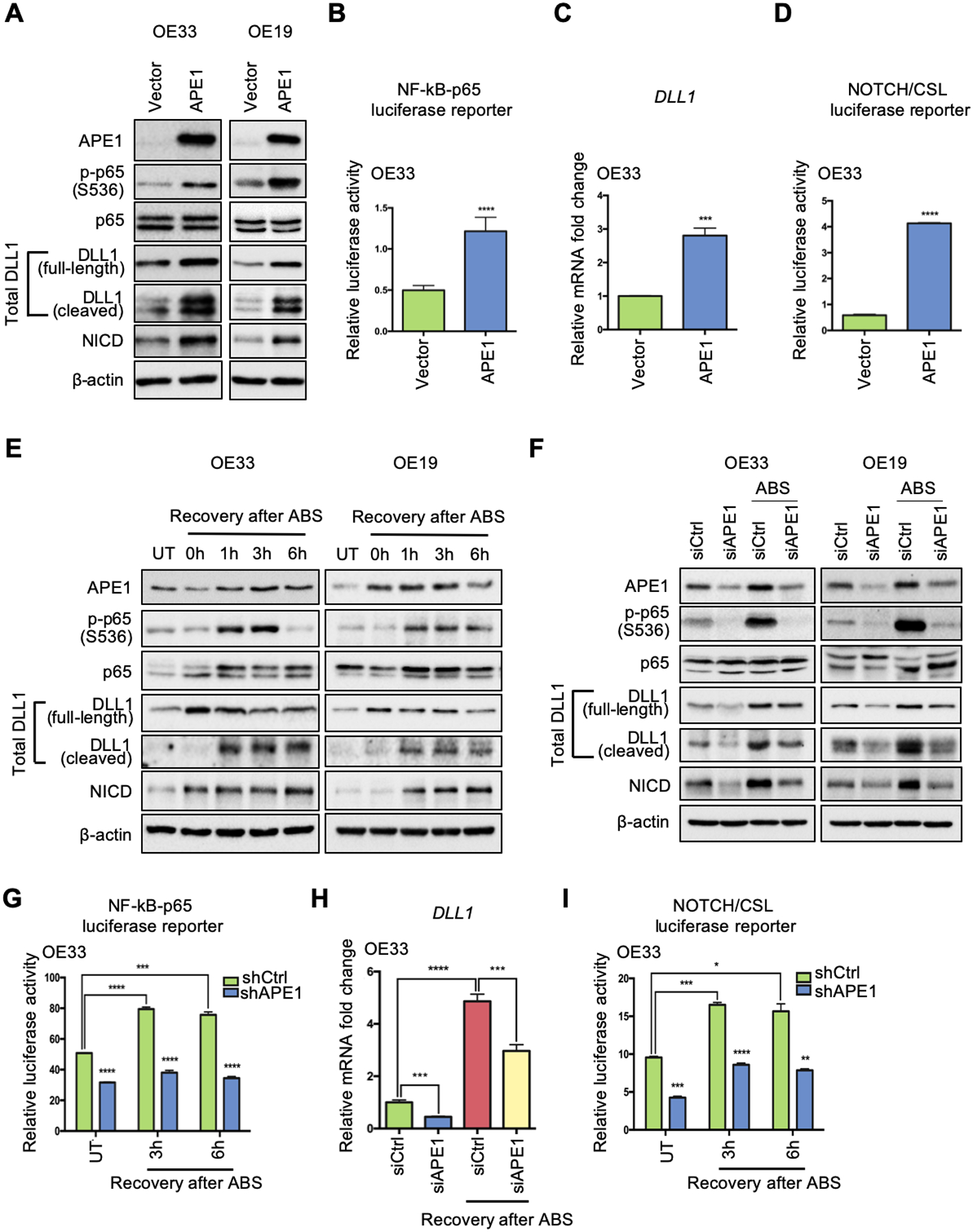
APE1 plays an essential role in ABS-induced NF-κB-DLL1-NOTCH activation. (A) Western blots were used to detect protein levels of interested genes after ectopic APE1 overexpression in OE33 (left) and OE19 (right) cells. (B) NF-κB-p65 luciferase activity was examined after APE1 overexpression. (C) qRT-PCR for DLL1 mRNA expression was performed in the condition of APE1 overexpression. (D) NOTCH/CSL luciferase reporter assay was performed to confirm NOTCH transcriptional activity after APE1 overexpression. (E) Western blots were used for detecting protein expression of APE1, phosphor-p65, total p65, total DLL1, NICD and β-actin at different time points of recovery from ABS exposure in OE33 (left) and OE19 (right) cells. (F) OE33 (left) and OE19 (right) cells were transfected with APE1 siRNA (siAPE1) or control siRNA (siCtrl) before ABS exposure; protein levels of interested genes were examined by Western blot analysis. (G) OE33 cells with stable APE1 knockdown (shAPE1) or control (shCtrl) were exposed to ABS, NF-κB-p65 activity luciferase reporter assay was performed at 3h and 6h recovery time points after ABS exposure. (H) APE1 knockdown (siAPE1) blocked ABS-induced DLL1 upregulation in OE33 as comparing to control cells (siCtrl). (I) NOTCH/CSL luciferase activity was checked in APE1-knockdown (shAPE1) and scramble shRNA control (shCtrl) OE33 cells with or without ABS exposure. Statistical data are shown as mean ± SEM. *p<0.05, **p<0.01, ***p<0.001 and ****p<0.0001 as calculated by t test for two group comparisons.