Figure 6.

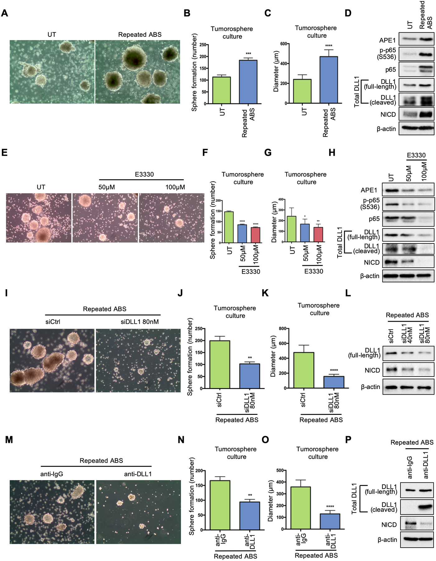
EAC tumorosphere formation is enhanced in response to acidic bile salts. (A) OE33 cells were exposed to ABS (200uM, pH5.5) 20min per day for 14 days. Then the cells were seeded for 3D tumorosphere culture. Untreated OE33 cells (UT) were used as a control. Representative images of the OE33-derived tumorospheres under white light. (B) and (C) Quantification of number (B) and size (C) of the tumorospheres in A. (D) Western blots of APE1, phosphor-p65, total p65, total DLL1(full-length and cleaved form), NICD and β-actin in the tumorospheres with and without repeated ABS exposure. (E) Tumorospheres derived from untreated OE33 cells were incubated with or without indicated doses of E3330 during culture; representative images of the OE33-derived tumorospheres with E3330 under white light. (F) and (G) Quantification of number (F) and size (G) of the tumorospheres in E. (H) Western blots of APE1, phosphor-p65, total p65, total DLL1(full-length and cleaved form), NICD and β-actin in the tumorospheres with E3330. (I) Tumorospheres derived from repeated-ABS-treated OE33 cells were transfected with DLL1 siRNA (siDLL1) or scrambled control (siCtrl); representative images of the tumorospheres with or without transient knockdown of DLL1 under white light. (J) and (K) Quantification of number (J) and size (K) of the tumorospheres in I. (L) Western blots of full-length DLL1, NICD and β-actin in the tumorospheres with and without indicated concentration of DLL1 siRNA. (M) Tumorospheres derived from repeated-ABS-treated OE33 cells were incubated with DLL1 neutralizing antibody or anti-IgG; representative images of the tumorospheres with DLL1 neutralization under white light. (N) and (O) Quantification of number (N) and size (O) of the tumorospheres in M. (P) Western blots of total DLL1 (full-length and cleaved form), NICD and β-actin in the tumorospheres with DLL1 neutralizing antibody or anti-IgG. Statistical data are shown as mean ± SEM. *p<0.05, **p<0.01, ***p<0.001 and ****p<0.0001 as calculated by t test for two group comparisons.
