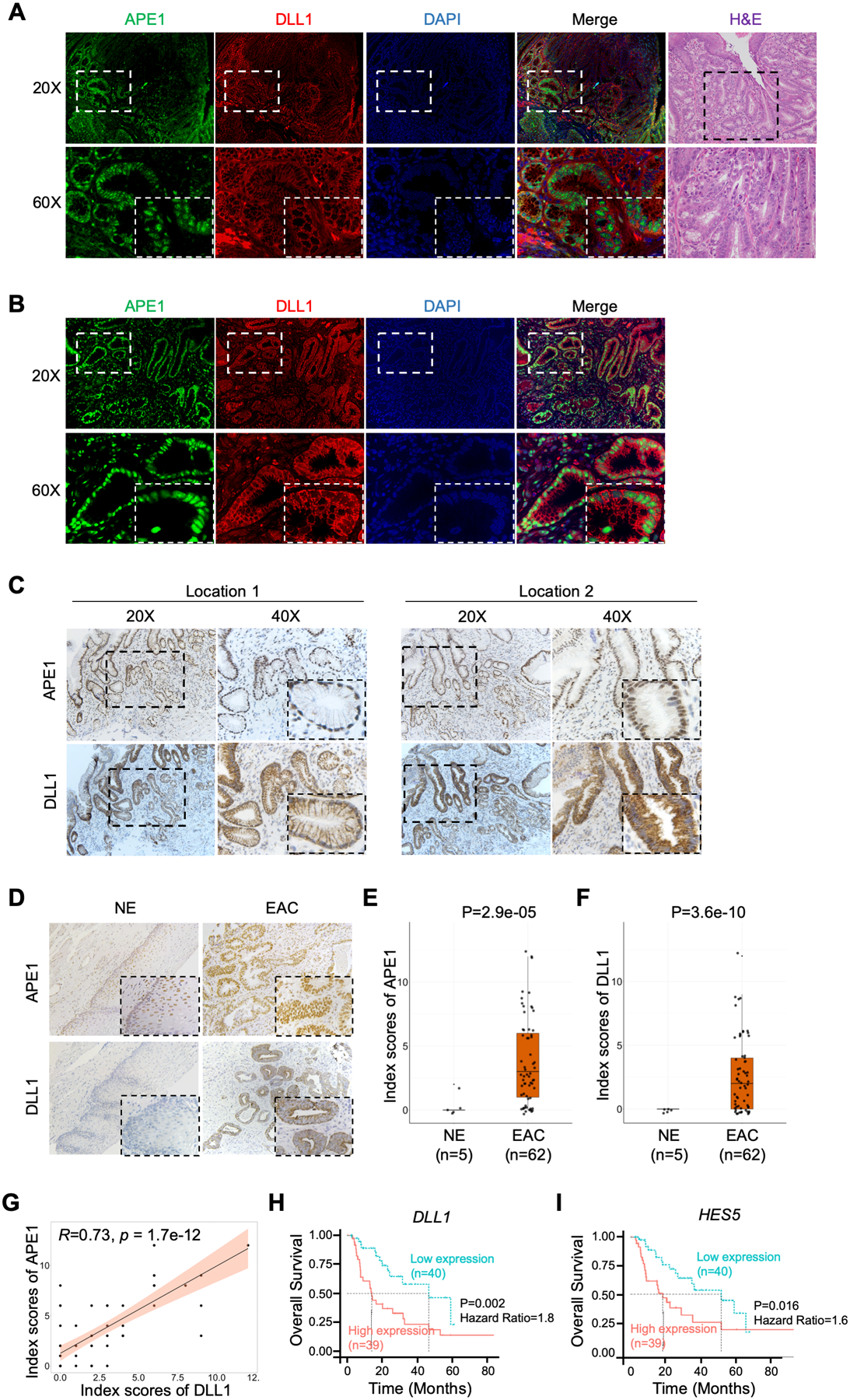
Figure 7.

DLL1 is upregulated in neoplastic mouse and human esophageal tissues and correlates with poor prognostic outcomes in human EAC. (A) and (B) Representative immunofluorescent staining images of APE1 (green) and DLL1 (red) in EAC tissue samples from pL2-IL1ß transgenic mouse (A) and human EAC tissue (B). DAPI was used for nuclear staining. (C) and (D) Representative IHC staining images of APE1 and DLL1 using the slides of the same human EAC tissue as B (C) and human EAC tissue microarrays (TMA) (D). (E) and (F) Comparisons of IHC index scores of APE1 (E) and DLL1 (F) between normal esophageal epithelium (n=5) and EACs (n=62) in human EAC TMA were shown. (G) Pearson correlation analysis of the IHC index scores of APE1 and DLL1 protein from human EAC TMA. (H) and (I) Kaplan-Meier plots were used for the survival analysis in the TCGA-EAC database, comparing DLL1-high patients with DLL1-low patients (H), or comparing HES5-high patients with HES5-low patients (I).
