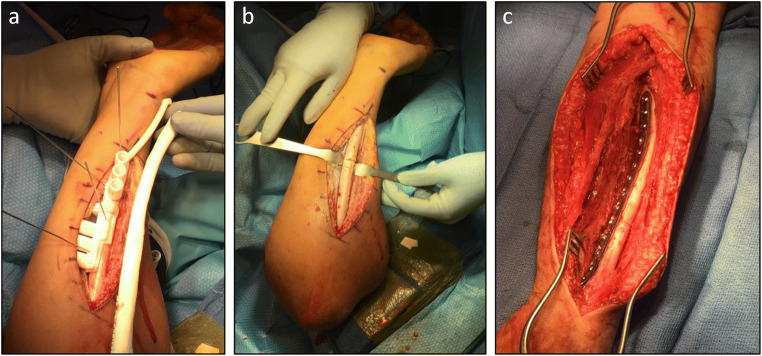
Fig. 5.
Intraoperative photographs demonstrating osteotomy using patient-specific cutting guide for patient in Fig. 2. a Patient-specific ulnar cutting guide held to patient’s ulna by wires; a plastic model of patient’s ulna can be used as an additional confirmation of guide placement accuracy. b Ulnar drill holes and osteotomy prior to reduction and internal fixation. c Volar view of internally fixed two-level radial osteotomy. (Courtesy of Sanjeev Sabharwal, MD, MPH)

