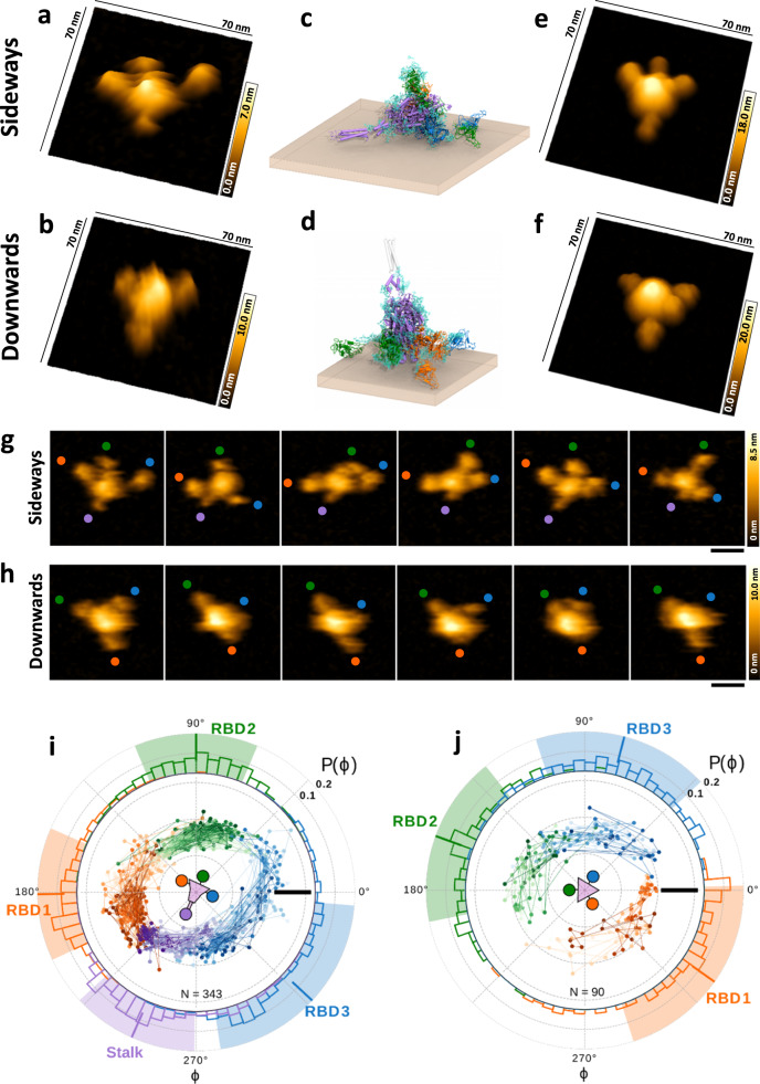
Fig. 1. High-speed AFM analyses demonstrating the high structural flexibility of the SARS-CoV-2 Spike trimer.
a, b 3D AFM image of the Spike trimer laying on mica sideways (a) or downwards (b). c, d Spike trimer model, corresponding to the conformation experimentally observed in (a) and (b), respectively. S1 domains with three RBDs are shown in orange, green, and blue. The S2 domain including the stalk is colored in purple. Glycans are shown as cyan sticks and the brown sheet represents the mica surface. The stalk is invisible in the downward configuration (b) and colored in faded gray (d). e, f 3D simulated AFM images based on the MDS models from (c) and (d). Snapshots from a movie of a single Spike trimer laying sideways (g) or downwards (h) on mica. The colored circles follow the position of the stalk (purple), and each of the RBDs (orange, green, and blue) over time. i, j Trajectories of stalk and RBDs in polar representation captured in movies (Supplementary Movies 1 and 2), corresponding to the sequences shown in (g) and (h). Radial coordinates denote the distance from the Spike center, ɸ is the rotational angle. Lines with increasing color intensity follow the trajectories of structures over time. The stalk is shown in purple, RBD1 in orange, RBD2 in green, and RBD3 in blue. Outer ring: The distribution of ɸ is shown in the colored histograms. Average values of ɸ are marked by a bar and the respective standard deviations by a background field in the same color. Spike orientation is schematically shown in the center. Images in a, b, g, and h were captured at a scanning speed of 154 ms/frame. Scale bar in g and h: 20 nm. Scale bar in i and j: 10 nm. Source data are provided as a Source Data file.

