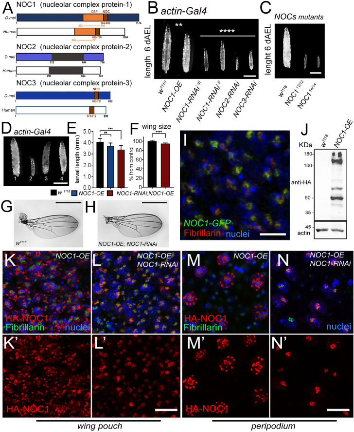
Fig. 1.
NOC1 is expressed in the nucleolus and its reduction, as for NOC2 and NOC3, affects animal growth and survival. (A) Schematic representation of Drosophila NOC1, NOC2 and NOC3 proteins and their human homologs, called CEBPz, NOC2L and NOC3L, respectively. NOC1 protein contains a CBP domain (CCAAT-binding domain), in orange, that shares 32% identity between sequences. The conserved NOC domain of 45 amino acids, present only in NOC1 and NOC3, is presented in brown; this shares 48% and 38% sequence identity between Drosophila and human proteins, respectively. NOC2 protein shares an overall 36% identity between Drosophila and human proteins; black represents the region of highest conservation (48%). (B) Photos of third-instar larvae expressing the indicated transgenes under the actin driver, taken at 120 h AEL. (C) Photos of control and NOC112 and NOC114 mutant third-instar larvae of 120 h AEL. (D) Photographs of larvae at 120 h AEL expressing the following transgenes (1) control w1118, (2) NOC1-RNAi, (3) NOC1 overexpression (OE), (4) NOC1-RNAi; NOC1-OE using the actin-Gal4 driver. (E) Larval length measured in mm at 120 h AEL. (F) Quantification of wing area/size in animals of the indicated genotype; the number is expressed as mean±s.d. percentage of the control actin-w1118, set at 100%. For E and F, at least 10 animals were used for each genotype; the experiment was repeated twice. (G-H) Photos representing wings from females of the indicated genotypes. (I) Confocal image of cells from the wing imaginal disc showing NOC1–GFP expression visualized using anti-GFP antibodies in green and anti-fibrillarin in red; nuclei are visualized with Hoechst. (J) Western blot from larval lysates expressing HA-NOC1 under the actin promoter. A band of ∼130 kDa is the expected size for NOC1, is visualized by anti-HA antibody with a few other bands at lower molecular mass; actin is used as control loading. (K–N) Confocal pictures of cells from the wing imaginal discs (K,L) or from the peripodial epithelium (M,N) expressing HA-NOC1 (K–M) or HA-NOC1;NOC1-RNAi (L,N) using the engrailed promoter. NOC1 expression was visualized using anti-HA antibodies in red and anti-fibrillarin in green. NOC1 expression alone is shown in K′–N′. **P<0.01; ***P<0.001; ****P<0.0001 [one-way ANOVA with Tukey multi-comparisons test (E); unpaired two-tailed Student's t-test (F)]. Images and blots shown are representative of three experiments. Scale bars: 1 mm (B–D,G,H); 5 µm (I), 10 μm (L,N).

