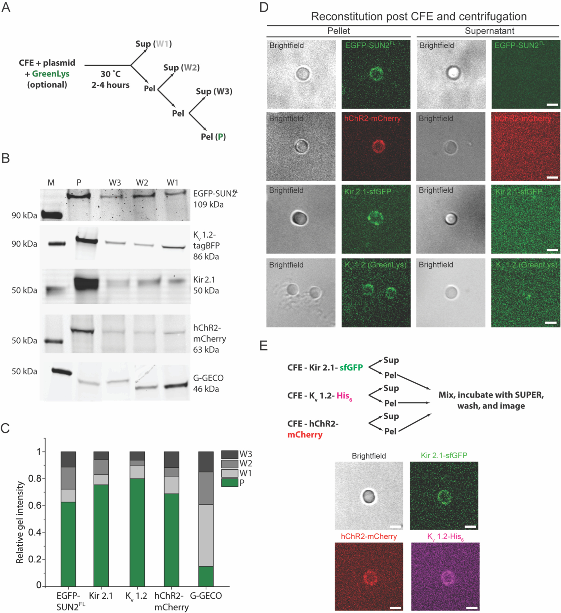
Figure 5. Direct reconstitution of FL membrane proteins mediated by ER-derived microsome fusion is used for the reconstitution of different or multiple membrane proteins.

A) Illustration of the experimental workflow used in panels B-E of this figure. B) Representative in-gel fluorescence images of the pellet and supernatant fractions from the microsome enrichment experiment depicted in panel A for the indicated CFE synthesized and GreenLys-labeled protein constructs. Lane M indicates the protein ladder. C) Bar graph of the normalized band intensities quantified from the in-gel fluorescence images presented in panel B. For each protein, band intensities were normalized by the sum of intensities in all lanes for the corresponding protein. D) Representative brightfield and confocal fluorescence images of SUPER templates incubated in and isolated from the pellet (denoted as P in panel A) or the supernatant fraction from the first wash (denoted as W1 in panel A) from microsome enrichment experiments where the indicated membrane protein constructs were synthesized. Scale bars: 5 μm. E) Experimental design for multiple membrane protein reconstitution (top) and representative confocal fluorescence images of SUPER templates incubated with microsome-enriched fractions of completed CFE reactions that synthesized the indicated proteins (bottom). Scale bars: 5 μm.
