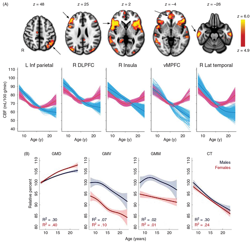
Figure 4.

(A) After reaching its peak level in preadolescents, cerebral blood flow (CBF) declines in male adolescents toward levels seen in young male adults. In females, CBF also declines in early adolescence but then increases in specific regions in late adolescence. These regions are denoted by arrows on the MR images; L Inf Parietal, left inferior parietal cortex; R DLPFC, right dorsolateral prefrontal cortex; R insula, right insula cortex; vMPFC, ventromedial prefrontal cortex; R Lat Temporal, right lateral temporal cortex. Source: Reused, with permission, from Satterthwaite TD, et al., 2014 (388). © 2014, National Academy of Sciences. Figure 2 from https://www.pnas.org/content/111/23/8643. (B) Structural changes in cerebral cortex during adolescence. Gray matter density (GMD), determined from T1 MRI intensity averaged over the entire brain, increases in adolescence, while gray matter volume (GMV), gray matter mass (GMM=GMD × GMV), and cortical thickness (CT) largely decrease in males and females. Note that the higher CBF per unit mass in females at the end of adolescence is associated with higher GMD and is distributed to a lower gray matter volume and mass in females compared to males, thereby attenuating sex differences in volumetric blood flow to the entire cortical volume. Values are plotted as percentages of the fitted value for males at 8 years of age. Shaded bands correspond to two SE of the fit. Source: Adapted, with permission, from Gennatas ED et al., (156).
