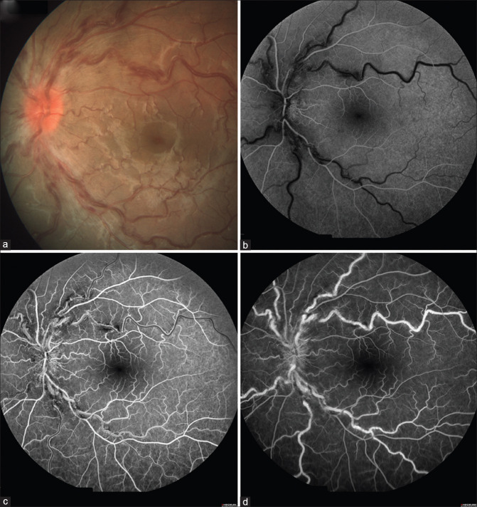
Figure 2.
Color photograph of the left eye showing (a) hyperemic, swollen optic disk with tortuous and engorged retinal veins with scattered retinal hemorrhages. Fundus fluorescein angiography of the left eye (b, c, and d) showing a delay in venous filling in the early phase of the angiogram and blocked fluorescence corresponding to the areas of hemorrhages

