Figure 1.

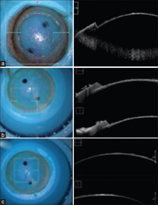
Microscopic and OCT imaging. Selected OCT images from three different corneas cut with VisuMax femtosecond laser. OCT = optical coherence tomography

Microscopic and OCT imaging. Selected OCT images from three different corneas cut with VisuMax femtosecond laser. OCT = optical coherence tomography