
Figure 1.
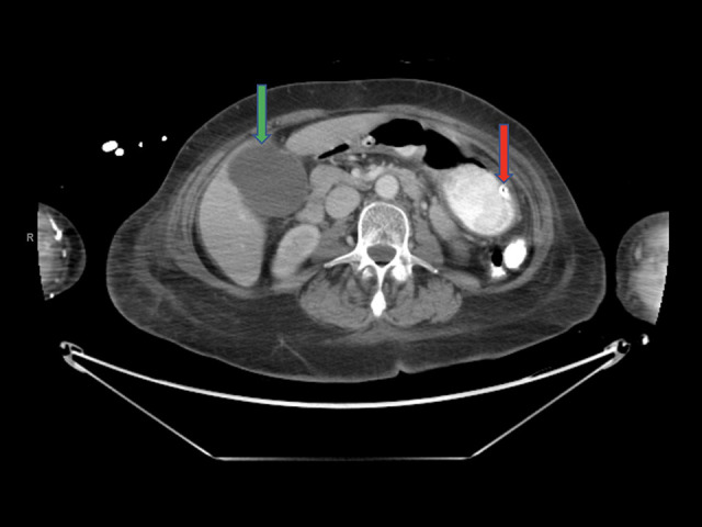
CT abdomen and pelvis with i.v. contrast and oral Gastrografin (GE Lightspeed RT 16). A CT obtained on day 3 of hospitalization demonstrated a nasogastric tube with its tip in the stomach (red arrow), moderately distended gallbladder with mild biliary ductal distention (green arrow), and no bezoar identified.