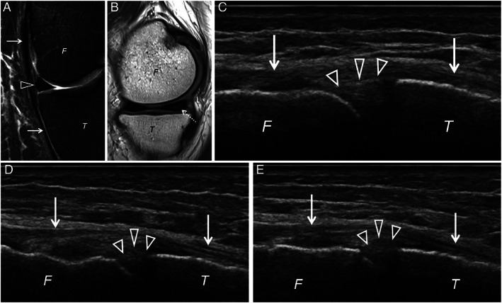
Figure 3.

Left knee of a 65‐year‐old male showing the medial meniscus (arrowhead) on (A) coronal PDW fat‐saturated at the level of the medial collateral ligament (arrows) without extrusion. Sagittal PDW magnetic resonance (MR) imaging (B) shows a mucoid degeneration (dashed arrow) of the posterior horn of the medial meniscus. Supine relaxed ultrasound (C) of the medial meniscus (arrowheads) at the level of the medial collateral ligament shows no extrusion. However, supine stressed ultrasound (D) and especially weight‐bearing ultrasound (E) shows a mild meniscal extrusion. F, femur; T, tibia.
