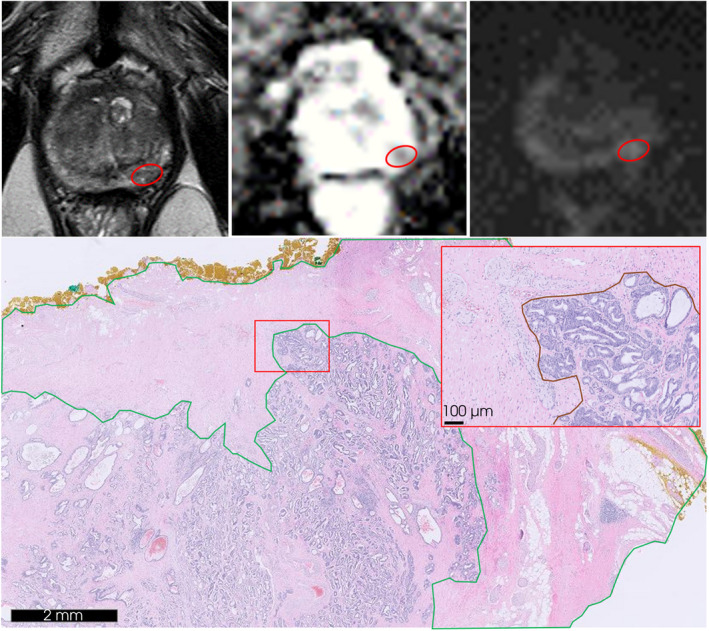
Fig. 2.

An example of in‐field recurrence at the left mid‐apex posterolateral peripheral zone on follow‐up MRI and corresponding haematoxylin and eosin stained (H&E) slide of the resected prostate specimen. Red circle demonstrates the aberrant lesion of 8 mm on MRI series: transverse T2‐weighted (upper left), apparent diffusion coefficient (upper mid) and diffusion weighted imaging of calculated b‐values (upper right). H&E slide obtained from the corresponding prostate specimen tissue (down) displaying IRE treatment‐induced inflammation and fibrosis (green) and recurrent disease of adenocarcinoma Gleason score 3 + 4 = 7 (down right; red).
