
FIGURE 8.
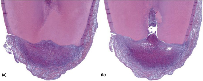
External inflammatory resorption. (a) Mandibular second premolar extracted with the periapical lesion attached. This section, which did not pass through the canal, shows extensive apical resorption (H&E stain; original magnification ×25). (b) Section taken approximately 120 sections away encompasses the apical foramen. In addition to resorption, the opposite phenomenon can be observed; that is, a large calcification partly embedded in the right apical dentin wall (original magnification ×25). (Reprinted from Pathways of the Pulp (12th Edition), Patel S, Durack C, Riccuci D, Bakhash A, Root Resorption, Copyright (2020) with permission from Elsevier)