
FIGURE 10.
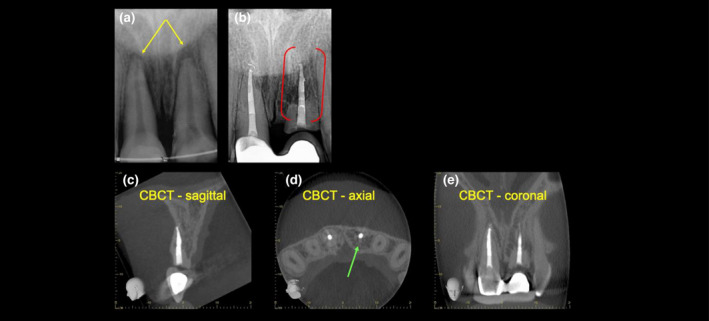
External replacement resorption. (a) A periapical radiograph of the maxillary central incisors with late presentation following severe luxation injury and complicated crown root fracture of the maxillary left central incisor. Note periapical radiolucency and blunting of the root ends (yellow arrows). (b) A 5‐year review radiograph following RCT of both teeth reveals direct bone replacement of the root dentine on the maxillary left central incisor (red brackets). Note the left central incisor has been decoronated and a restored with a temporary resin‐bonded bridge. Note the periapical radiolucency on both teeth have healed. (c–e) Sagittal, axial and coronal CBCT slices through the same tooth confirm an almost complete bony replacement of root dentine associated with ERR (green arrows). Reprinted from British Dental Journal, Volume 224, Patel S, Saberi N: The ins and out of root resorption. 691–699 Copyright (2018) with permission from Springer Nature