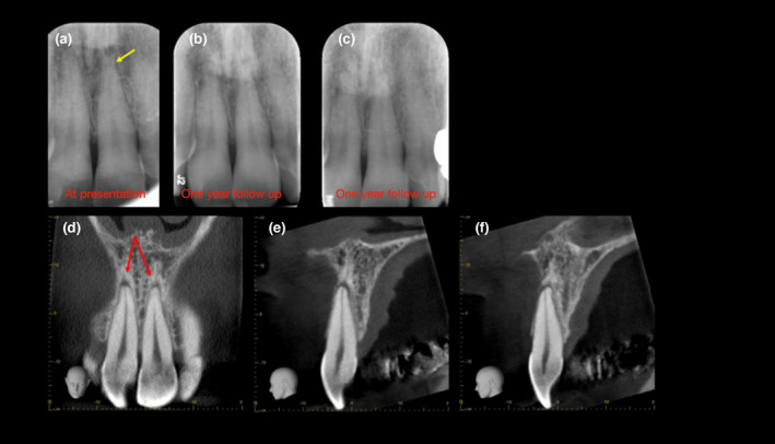
FIGURE 11.

Transient apical breakdown (a) A periapical radiograph revealing widening of the periodontal ligament space and loss of apical lamina dura following subluxation injury to the maxillary anterior teeth (yellow arrow). (b) One‐year follow‐up radiograph demonstrates an improvement, and (c) a 10‐degree vertical shift eliminates the presence of the lesion altogether, confirming a diagnosis of transient apical breakdown. (d–f) Coronal and sagittal CBCT slices of the same teeth, however, reveal well‐defined periapical radiolucencies associated with the maxillary right and left central incisors (red arrows); therefore, the diagnosis was changed to chronic periapical periodontitis associated with infected necrotic pulp. This case highlights anatomical noise and subtle geometrical distortion associated with conventional radiographs, which may be overcome with CBCT (Reprinted from British Dental Journal, Volume 224, Patel S, Saberi N: The ins and out of root resorption. 691–699 Copyright (2018) with permission from Springer Nature)
