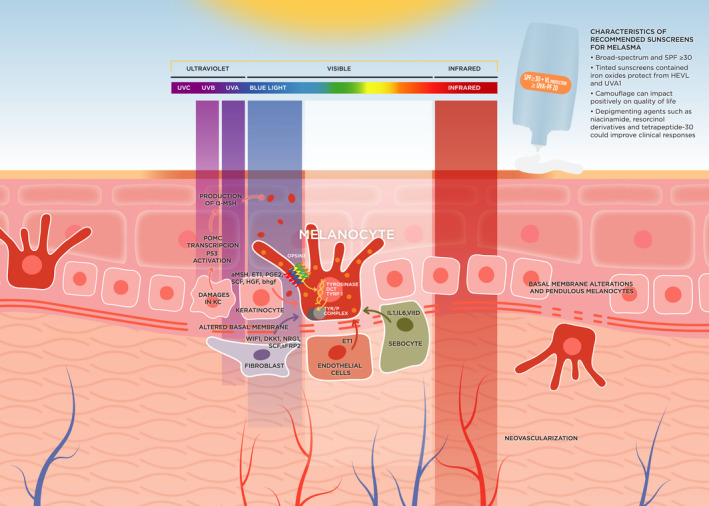
FIGURE 1.

Diagram summarizing pathologic changes and mechanisms in melasma. Melanocytes are producing and distributing melanin to surrounding keratinocytes. UVB‐induced DNA damage of keratinocytes activates P53, binding to the POMC promoter and finally leads to the secretion of α‐MSH by keratinocytes. Chronic exposure to UVA and VL alters the dermal component. VL induces skin pigmentation through the activation of Opsin 3, a photoreceptor, which mediates the expression and activity of the rate‐limiting enzyme tyrosinase in melanocytes. Basal membrane alteration promotes the descent of melanocytes to the dermis (pendulous melanocytes). Keratinocytes produce cytokines and hormones, especially after UVB exposure. Fibroblasts secrete factors that influence melanogenesis and melanocyte proliferation. The increased vascularization plays a key role, especially with endothelial cells that produce ET1, which is a potent activator of melanogenesis. Abbreviations: DCT dopachrome tautomerase; DKK1, Dickkopf 1; DNA, Deoxyribonucleic Acid; ET1, Endothelin 1; HGF, hepatocyte growth factor; KC, Keratinocytes; p53, Tumor protein p53; PGE2, prostaglandin‐E2; POMC, Proopiomelanocortin; SCF, stem cell factor; sFRP2, Frizzled‐related protein 2; SPF, Sun Protection Factor; TYR/P COMPLEX, tyrosinase‐related protein complex; TYRP1, tyrosinase‐related protein 1; UVA, Ultraviolet A; UVB, Ultraviolet B; UVC, Ultraviolet C; WIF1, Wnt inhibitory factor 1; α‐MSH, α‐Melanocyte‐stimulating hormone
