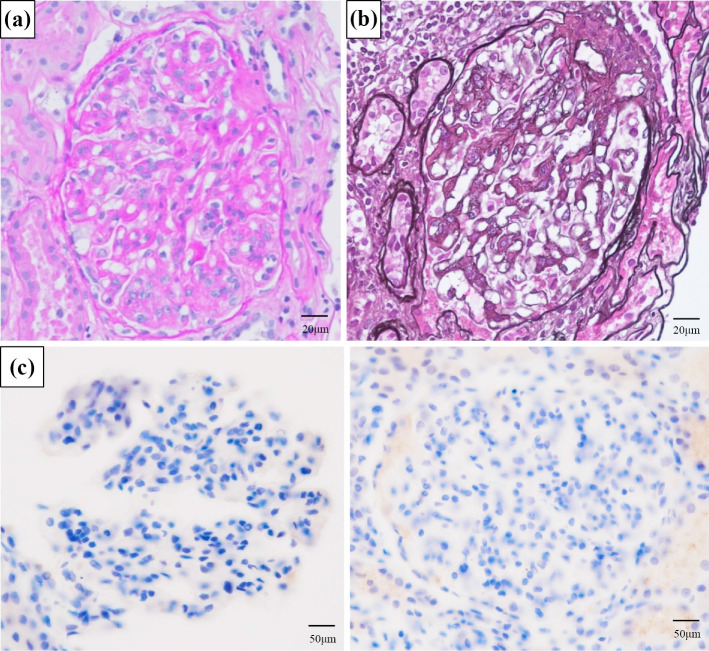
Figure 1.
Light microscopy revealed diffuse mesangial proliferative nephritis with subendothelial deposition. (a) Periodic acid-Schiff stain. (b) Periodic acid methenamine stain. (c) Granular deposition in kappa and lambda light chain staining was negative in the glomerular tuft mesangial region and partial endothelium.

