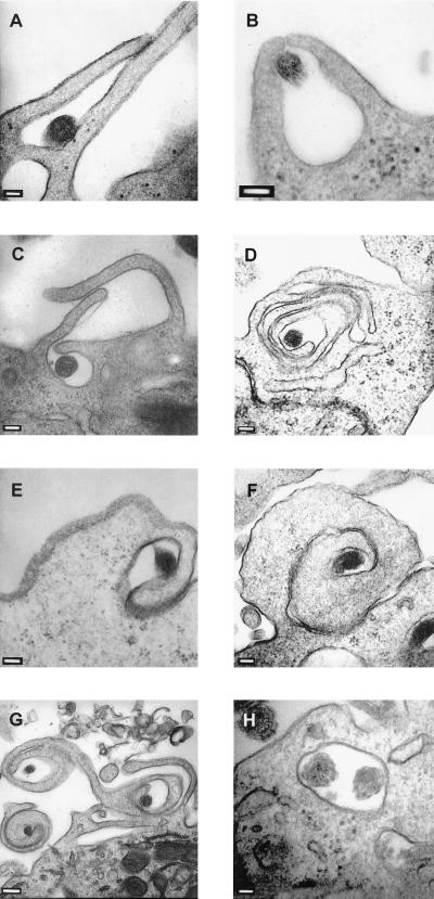
FIG. 3.
Phagocytosis of T. pallidum by XS52 cells. T. pallidum cells were incubated with XS52 cells at a ratio of 1,000 spirochetes/cell for 24 h. Samples were prepared for thin-section (80 nm) TEM and negatively stained with lead citrate and uranyl acetate. (A and B) Two examples of conventional phagocytosis. (C) Overlapping coiling phagocytosis. (D) Overlapping coiling phagocytosis in the process of internalizing T. pallidum. (E) The initial pseudopod of rotating coiling phagocytosis surrounding a spirochete. (F) A pseudopod whorl of rotating coiling phagocytosis. (G) Three pseudopod whorls of rotating coiling phagocytosis. (H) Two organisms internalized in what appears to be a membrane-bound vacuole. Bars: 100 (A to F) 200 (G), and 50 nm (H).

