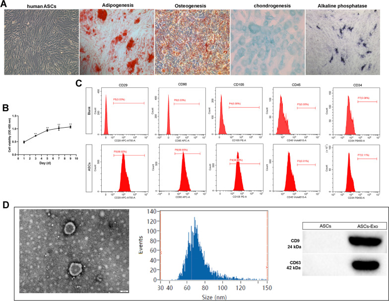
Fig. 1.
Classification of adipose stem cells (ASCs) and exosomes. A ASCs exhibited a representative spindle-like morphology (scale bar: 100 μm). ASCs were stained with Oil red O, Alizarin red, alkaline phosphatase, and Alcian blue stain. B The cell proliferation of ASCs was tested by CCK-8 assay. **P < 0.01 (VS. 1 day). C Flow cytometric analysis of characteristic cell surface markers of ASCs (CD29, CD90, CD105, CD45, and CD34). D Morphology of exosomes observed by transmission electron microscopy (TEM). Scale bar: 100 nm. Particle size distribution of exosomes was measured by Nanosight. The expression of exosome surface markers (CD9 and CD63) was measured using western blot

