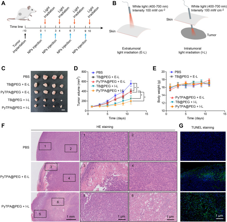
Figure 1.
(A) The objective to construct Hela subcutaneous tumor and the treatment process. (B) Schematic illustrating treatment by extratumoral light irradiation (E-L) or intratumoral light irradiation (I-L). Light intensity:100 mW cm−2, time: 20 min. (C) Representative tumor images of the different groups after treatment. (D) Growth kinetic curves of tumors in tumor-bearing mice after receiving PyTPA@PEG or TB@PEG -mediated extratumoral light irradiation (E-L) or intratumoral light irradiation (I-L). The data were reported as mean ± SD (n = 4) and analyzed by unpaired t-test. * p< 0.05. (E) Body weight changes in mice during treatment. (F) The histological changes of tumors were observed by H&E staining. (G) Cell apoptosis of tumors were detected by TUNEL staining.

