Figure 3.
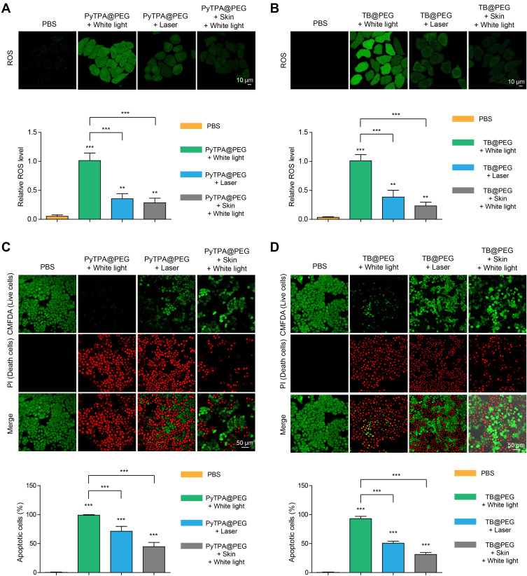
In vitro cell experiments. (A) Detection of intracellular ROS generation by DCFH-DA in HeLa cells after incubation with PyTPA@PEG NPs (10 µM), followed by white light, 532 nm laser irradiation and white light penetration of skin (all light intensity: 200 mW cm−2, time: 3min). (B) Detection of intracellular ROS generation after incubation with TB@PEG NPs (10 µM), followed by light (all light intensity: 200 mW cm−2, time: 5 min). (C) Hela cells were cultured with PyTPA@PEG NPs (20 µM) and then irradiated with white light, 532 nm laser irradiation and white light penetration of skin (all light intensity: 200 mW cm−2, time: 3 min). CMFDA: Ex = 488 nm; Em = 540 nm. PI: Ex = 543 nm; Em = 650 nm. (D) Hela cells were incubated with TB@PEG NPs (30 µM) and treatment with different light irradiation (all light intensity: 200 mW cm−2, time: 5 min). Data are represented as mean ± standard deviation (SD) and analyzed by two-sided Student’s t-test. **p < 0.01, ***p < 0.001.
Abbreviation: n.s., not significant.

