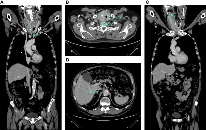
Figure 2.
axial (A) and coronal (B) CT-scan showing large inhomogeneous tumor with calcifications of the right thyroid lobe with left tracheal displacement, infiltrating the trachea and esophagus; confluent neck lymph node metastases (maximum diameter of 58 mm) (C); hepatic lesion of 12 mm at V segment (D).

