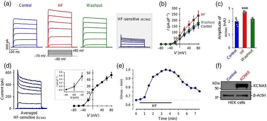
FIGURE 2.
Halofuginone (HF) reversibly increases whole-cell K+ currents in HEK cells transiently transfected with the human KCNA5 gene. (a) Representative outward K+ currents, elicited by depolarizing a cell from a holding potential of −70 mV to a series of test potentials ranging from −80 to +80 mV in increment of 20 mV, before (control), during (HF) and after (washout) extracellular application of 1-μM halofuginone. The halofuginone -sensitive K+ currents through KCNA5 channels (IKCNA5) shown in inset are obtained by subtracting the currents recorded before halofuginone application (control) from the currents recorded during HF application (HF). (b) Averaged current–voltage (I–V) relationship curves (means ± SE, n = 8) for IKCNA5 recorded before (control), during (HF) and after (washout) halofuginone application. The halofuginone curve is significantly different from control and washout curves. (c) Amplitude of IKCNA5 at +80 mV under control, HF and washout conditions. Data shown are means ± SEM; n = 8 cells; for two of the 8 cells we were unable to record current after application of HF. *P < 0.05, significantly different from control and washout. (d) Averaged halofuginone -sensitive IKCNA5 (left panel) and its I–V curve (right panel). Inset: enlarged portion of the I–V curve showing the halofuginone -sensitive IKCNA5 at negative potentials (−80 to −40 mV). (e) Time course of halofuginone -induced effect on IKCNA5. Normalized IKCNA5 at +80 mV before, during and after extracellular application of 1-μM halofuginone. (f) Western bot analysis on KCNA5 in HEK cells transfected with an empty vector (control, n = 6 cells) or the human KCNA5 gene (KCNA5, n = 6 cells)

