FIGURE 3.
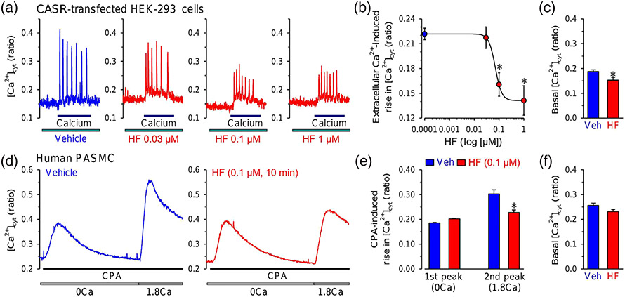
Halofuginone (HF) inhibits receptor-operated and store-operated Ca2+ entry. (a) Representative traces showing changes in [Ca2+]cyt before, during and after superfusion of extracellular application of 1.8-mM Ca2+ (calcium) in HEK-293 cells transfected with the calcium-sensing receptor (CaSR) gene (CASR) with (HF) or without (vehicle [Veh]) treatment of 0.03-, 0.1- and 1-μM halofuginone. (b) Summarized data showing the concentration–response curve of 1.8-mM Ca2+-induced increases in [Ca2+]cyt in CASR-transfected HEK-293 cells treated with different concentrations of halofuginone (0.0001 to 1 μM). Data shown are means ± SEM, n = 6 independent experiments for each data points. *P < 0.05, significantly different from 0.0001-μM halofuginone. (c) Summarized data (means ± SEM) showing the basal [Ca2+]cyt in CASR-transfected HEK-293 cells treated with Veh (n = 5) and all concentrations of halofuginone (0.03–1 μM) (HF, n = 7). *P < 0.05, significantly different from Veh. (d) Representative traces showing changes in [Ca2+]cyt before and during extracellular application of cyclopiazonic acid (CPA, 10 μM) in the absence (0Ca) or presence (1.8Ca) of 1.8-mM Ca2+ in human PASMCs shortly (10 min) treated with Veh (left panel) or 0.1-μM halofuginone (right panel). (e) Summarized data (means ± SEM, n = 6 independent experiments) showing the CPA-induced increases in [Ca2+]cyt in the absence (first peak, 0Ca) and presence (second peak, 1.8Ca) of 1.8-mM extracellular Ca2+ in human PASMCs treated with Veh or halofuginone (0.1 μM). *P < 0.05, significantly different from Veh. (f) Summarized data (means ± SEM) showing the basal [Ca2+]cyt in human PASMCs treated with Veh (n = 6 independent experiments) and 0.1-μM halofuginone (n = 6 independent experiments)

