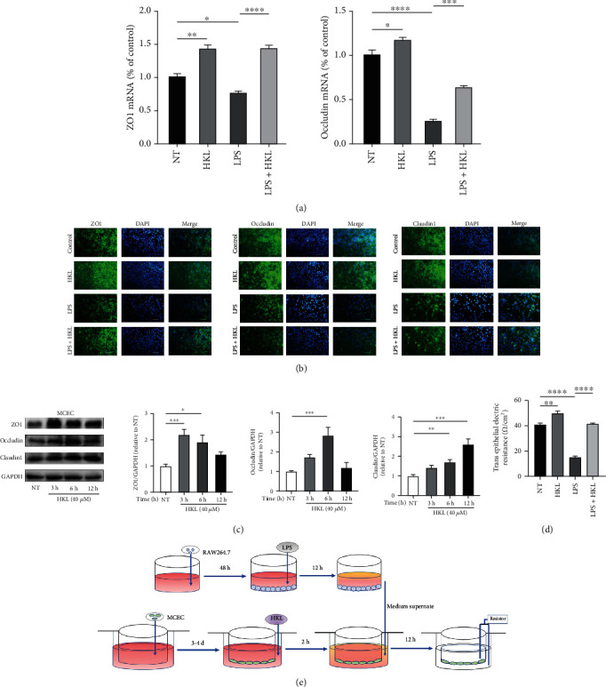
Figure 1.

HKL maintained a normal epithelium barrier in MCEC. (a) MCEC were pretreated with 40 μM HKL for one hour and then stimulated with the supernatant of 12h-LPS-induced RAW264.7 cells for 12 h. QRT-PCR analyses measured the mRNA expression of ZO1 and occludin; mRNA expression was normalized to β-actin expression. (b) Cell immunofluorescence of tight junction proteins in confluent MCEC pretreated with 40 μM HKL for one hour and then stimulated with the supernatant of 12h-LPS-induced RAW264.7 cells for 12 h. Fluorescent microscopy obtained representative images from three independent experiments after immunofluorescence staining of ZO-1, occludin, and Claudin1. DAPI staining was performed to identify nuclei. (c) MCEC were grown to confluence on 6-well plates and treated with HKL (40 μM) for the indicated periods. Western blots and related quantification of ZO-1, occludin, and Claudin1 are expressed as a percent of control. (d) TEER of various treatments of the MCEC monolayer. (e) Schematic diagram of the testing process of TEER. Data are expressed as mean ± SEM (n = 3). ∗P < 0.05, ∗∗P < 0.01, ∗∗∗P < 0.001, and ∗∗∗∗P < 0.0001.
