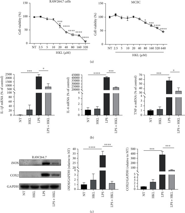
Figure 6.

HKL suppressed proinflammatory cytokine expression in LPS-stimulated macrophages. (a) CCK-8 analyzed the effects of HKL at different concentrations on the viability of RAW264.7 cells and MCEC. (b) RAW 264.7 cells were pretreated with 10 μM HKL for one hour and then stimulated with LPS (1 μg/ml) for 6 hours. QRT-PCR analyses of the mRNA expression of IL-1β, IL-6, and TNF-α were measured. mRNA expression was normalized to β-actin expression. (c) RAW 264.7 cells were pretreated with 10 μM HKL for one hour and then stimulated with LPS (1 μg/ml) for 12 h. Western blots and related quantification of iNOS and COX2 expressed as a percent of control. Data are expressed as mean ± SEM (n = 3). ∗P < 0.05, ∗∗∗P < 0.001, and ∗∗∗∗P < 0.0001.
