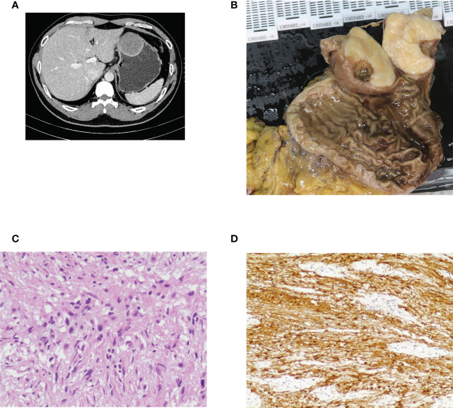
Figure 2.
Case illustration of gastric schwannoma. (A) Computed tomography showed a protruding lesion in the stomach. (B) The tumor was removed by surgical resection, and this was the resected tumor. (C) Histological results revealed spindle cell tumors. (D) Immunohistochemical staining of S100 was positive, consisting of schwannoma.

