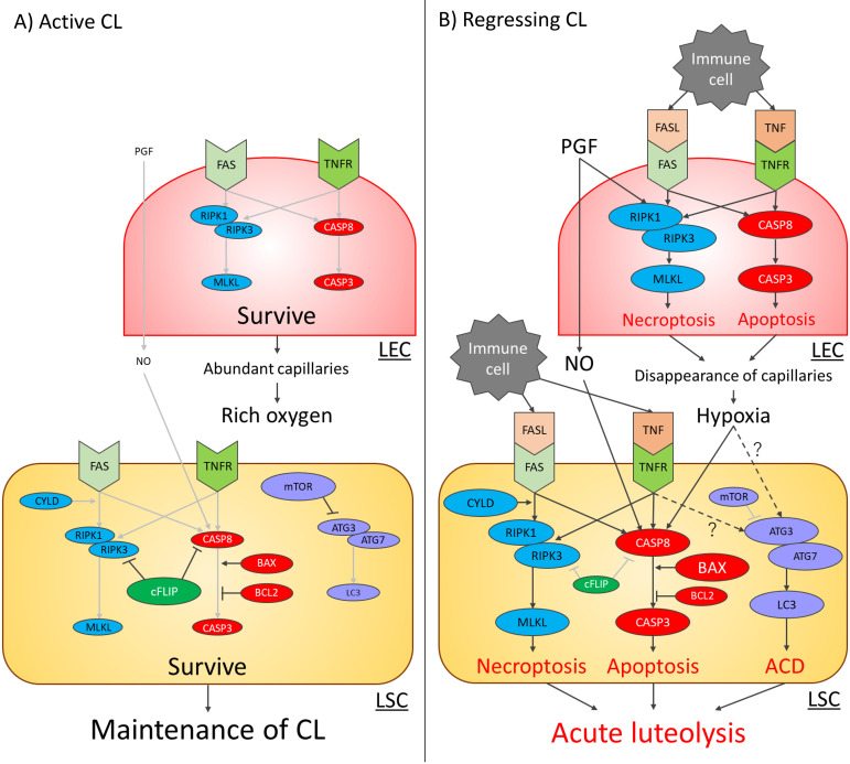
Fig. 1.
Programmed cell death (PCD) signaling pathways in bovine luteal endothelial cells (LECs) and luteal steroidogenic cells (LSCs) during the active CL (A) and regressing CL (B) stages. Red, purple, and light blue circles in the cells indicate apoptosis-related, autophagic cell death (ACD)-related, and necroptosis-related factors, respectively. In the active CL stage (A), the expression of extracellular PCD stimulators, including pro-inflammatory cytokines, immune cell-derived death ligands, and nitric oxide, is low. Thus, the expression of each PCD signal-related factor is also low, and these signals are weak (light gray arrows) in LECs and LSCs. Finally, both types of cells survive to maintain the CL. In the regressing CL stage (B), the expression of each PCD signal-related factor is stimulated by extracellular PCD stimulators and conditions of hypoxia. Therefore, each PCD signal becomes strong (black arrows), and PCD is induced. The details of the effects of the TNF-TNF receptor (TNFR) system and conditions of hypoxia on ACD-related signals (dotted arrows) are not well understood. Finally, interaction between various PCD results in structural luteolysis and the elimination of the active CL from the ovary.

