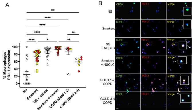
Figure 7.
Similar PD-L1 expression in AMs from patients with GOLD 1–2 COPD and patients with NSCLC. Immunofluorescence analysis of PD-L1 expression by bronchoalveolar lavage (BAL) AMs from Cohort 2. (A) empty symbols represent never-smokers (NS); yellow symbols represent subjects who stopped smoking > 1 year prior to the study; red symbols represent current smokers at the time of the study. (B) representative immunofluorescence pictures showing CD68+ (green, as a marker of macrophages) and PD-L1+ (red) cells in bronchoalveolar lavage from all the cohort 2 study subjects. Nuclear DNA was labeled with DAPI (blue). Confocal microscopy was performed with 60X and 100X objectives. The insets show CD68 + alveolar macrophages (AMs) that were positive (NS + NSCLC, smokers + NSCLC, and GOLD 1–2 COPD) or negative (NS, smokers, GOLD 3–4 COPD) for PD-L1.

