
Figure 7.
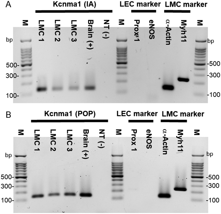
PCR to detect Kcnma1 (KCa1.1 α-subunit) in mouse lymphatic muscle cells (LMCs) isolated from inguinal-axillary (IA) lymphatic vessels (A) and popliteal (POP) lymphatic vessels (B). LMCs were collected by FACS analysis after isolation and digestion of lymphatic vessels from SMMHCCre;Rosa26mTmG mice. Kcnma1 showed strong bands in both LMC1 (168 cells), 2 (144 cells) and 3 (88 cells) from IA samples and LMC1 (76 cells), 2 (79 cells) and 3 (80 cells) from POP samples. A small amount of brain cDNA was used as a positive control (Lane 5). A representative panel of cell markers from LMC3 is shown. α-actin and Myh11 were detected but not Prox1 or eNOS, indicating no contamination by lymphatic endothelial cells (LEC) (Lane 8-11). Kcnma1 (135 bp), Prox1 (218 bp), eNOS (143 bp), α-actin (129 bp) and Myh11 (myosin-11; 238 bp). M = Marker; NT = negative; bp = base pairs.