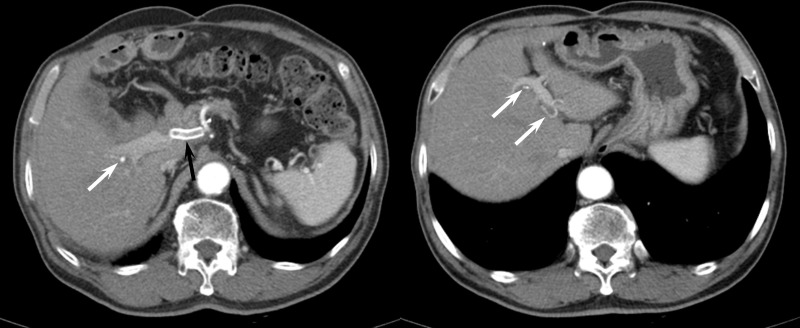
Figure 3.
A 71-year-old male who underwent covered stent placement for postoperative GDA pseudoaneurysm. (A) A contrast-enhanced CT obtained 22 months after stent placement shows thrombosed covered stent (black arrow). However, the right (white arrow) and left hepatic arterial flow (arrows in B) are maintained through collateral vessels.

