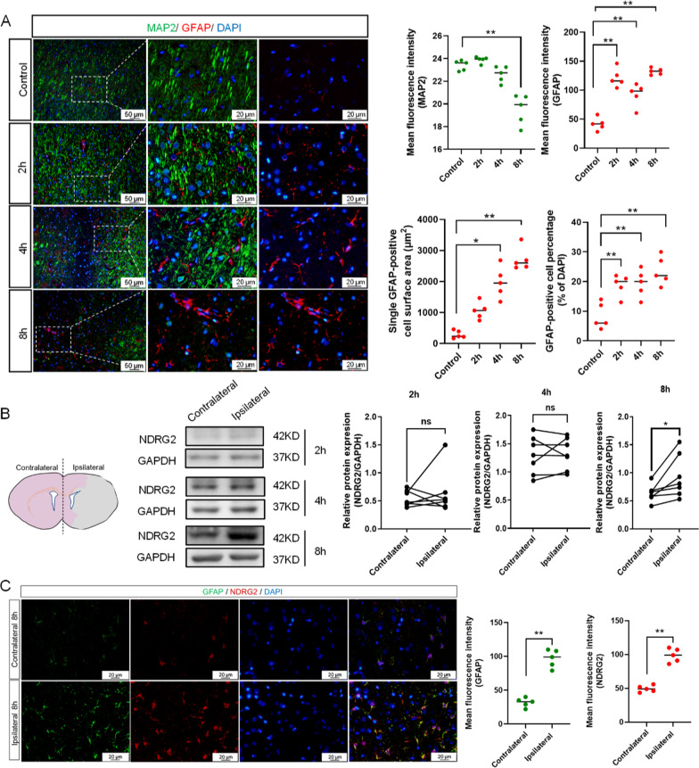
Fig. 1.
NDRG2 levels in reactive astrocytes are upregulated in ischemia. A Representative images of the cortex after 2 h, 4 h, and 8 h of middle cerebral artery occlusion (MCAO), double-stained for glial fibrillary acidic protein (GFAP, red) and microtubule-associated protein 2 (MAP2, green). Scale bar: 50 μm or 20 μm. Statistical significance was assessed using one‐way ANOVA followed by ordinary ANOVA or nonparametric test. Data are expressed as means ± standard deviations (SD); n = 5 random samples, *P < 0.05 and **P < 0.01 vs. Control. B Western blot analysis of NDRG2 expression in contralateral and ipsilateral brain tissues obtained 2 h, 4 h, and 8 h after MCAO surgery. Glyceraldehyde-3-phosphate dehydrogenase (GAPDH) is used as a loading control, with semi-quantification of western blot findings representing NDRG2 expression levels. Paired t-tests were used for statistical comparisons. Data are expressed as means ± SD. n = 7, ns = no significance and *P < 0.05 vs. Contralateral. C Representative brain sections obtained 8 h after MCAO surgery, double-stained for GFAP (green) and NDRG2 (red). Scale bar: 20 μm. Quantification of NDRG2 and GFAP expression is shown in the right panel. Unpaired t-tests are used for statistical comparisons. Data are presented as means ± SD. n = 5 random samples, **P < 0.01 vs. Contralateral

