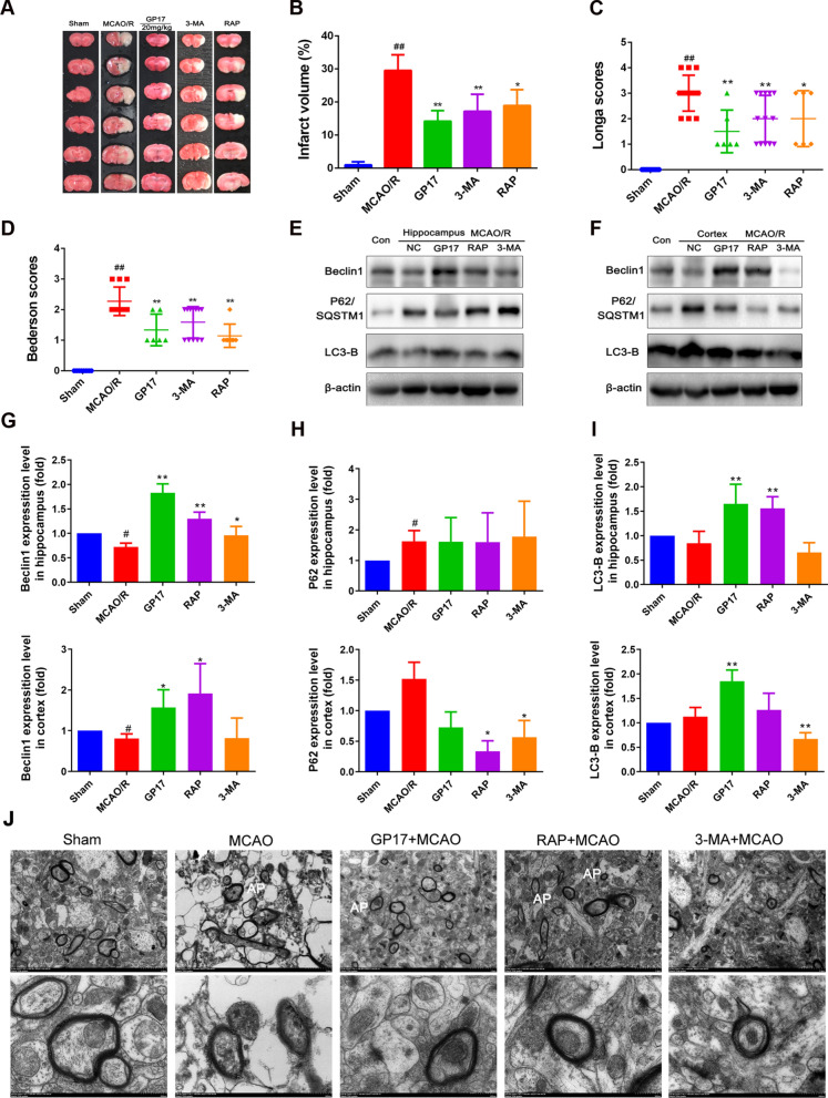
Fig. 2.
Effects of GP17, the autophagy activator RAP and the autophagy inhibitor 3-MA on infarct volume, neurological deficit scores, accumulation of autophagic vacuoles, and the Beclin1-LC3-P62 signalling pathway in rats with MCAO/R injury. A Representative images of TTC-stained brain sections from sham-operated or GP17-treated animals collected 24 h after infarction. Red tissue is healthy; white tissue is infarcted (n=3–6). B Quantitative analysis of the infarct volume (n=3). C and D. Neurological deficit scores in all groups (n=6–12). E and F The protein bands of LC3-B, Beclin1, and P62 in the ischemic brain sections were examined by western blot analysis. G–I. The relative expression levels of LC3, Beclin1, and P62 were quantified and analysed by using Gel-Pro analyser software (n=3–6). J. Accumulation of autophagic vacuoles (AP) was examined using a TEM; 5000, scale bars=2 μm, 20,000, scale bars=500 nm. Statistical comparisons were performed as follows: one-way ANOVA with Dunnett’s multiple comparisons test for (B, C, D and I); unpaired t test for (G and H); *P<0.05, **P<0.01 versus MCAO/R group; #P<0.05, ##P<0.01 versus Sham group

