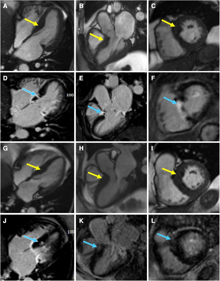
Figure 3.
Four-chamber (A, Supplementary material online, Video S1), three-chamber (B, Supplementary material online, Video S2), and basal short-axis cine steady-state free precession (SSFP) images (C, Supplementary material online, Video S3) of Twin A with corresponding delayed enhancement images (D–F) showing basal septal hypertrophy (arrows in A–C) with replacement fibrosis (arrows in D–F) in the basal septum consistent with infarct pattern after alcohol septal ablation. Four-chamber (G, Supplementary material online, Video S4), three-chamber (H, Supplementary material online, Video S5), and basal short-axis cine SSFP images (I, Supplementary material online, Video S6) of Twin B with corresponding delayed enhancement images (J–L) showing basal septal hypertrophy (arrows in G–I) with patchy mid myocardial replacement fibrosis (arrows in J–L) in the basal septum.

