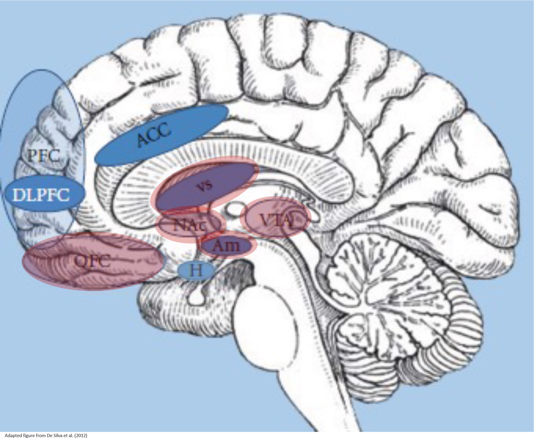
Figure 5: Brain circuits involved in the pathopsychology of BED.

Neuropsychological impairments of binge eating disorder (BED) have been explored in several brain imaging studies. The neurological basis of binge eating is composed of the hypothalamus (H) that regulates energy balance, such as food intake stimulated by gut hormones, the reward system that is representing motivational-affective functions (comprising the amygdala (Am), nucleus accumbens (Nac), ventral tegmental area (VTA), ventral striatum (VS) and orbitofrontal / ventromedial prefrontal cortex (OFC)), and cortical regions that are responsible for inhibitory control processes (comprising the prefrontal cortex (PFC), dorsolateral PFC (DLPFC), anterior cingulate cortex (ACC); insula and inferior frontal gyrus not shown). These three systems interact while binge eating episodes and mirror main components of impulsivity, that is, reward sensitivity and inhibitory control.
