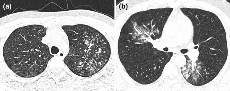
Fig. 4.
a A patient with APTB from an internal test set, male, 20 years old, weak for 1 month. Chest CT images showed multiple small nodules distributed along the bronchus in the upper lobe of the left lung, with blurred boundary and uneven thickening of some bronchial walls. The probability of diagnosing APTB output from the 3D-CNN optimal model was 1.000. b A patient with CAP from an external test set, male, 30 years old, fever for 6 days, cough and expectoration for 4 days. Chest CT images showed patchy high-density shadows in the middle lobe of the right lung and the lower lobe of the left lung, with blurred boundaries, and air bronchogram sign. The probability of diagnosing APTB output from the 3D-CNN optimal model was 0

