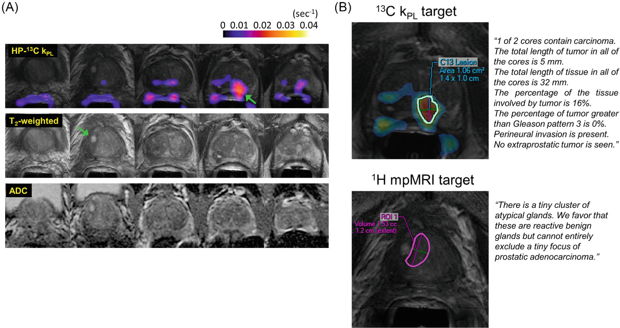
FIGURE 6.

(A) Shows an example of an integrated HP-13C research and standard 1H mpMRI study (patient 1) including key multiparametric T2-weighted, diffusion, and kPL images identifying the biopsy target. One 1H target (PIRADS 4) was identified at right mid-base transition zone and one 13C research target (kPL = 0.0378 s−1) at left mid-apex peripheral zone, as indicated by the arrows. (B) 13C and 1H mpMRI biopsy targets as drawn by an experienced abdominal radiologist. Pathological diagnosis of the tissue sample from the 13C target was Gleason 3+3 cancer (16% involvement, 1/2 cores), whereas that from the 1H-MRI target was described in the histology report as “rare atypical glands”
