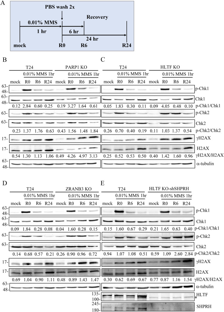
Fig 8. PARP1-, HLTF-, and ZRANB3-depleted cells show higher levels of γH2AX and checkpoint activation than control cells after MMS treatment.
(A) The protocol of DNA damage recovery assay. Cells were treated with 0.01% MMS for 1 hour (R0), followed by recovery in fresh medium for 6 (R6) and 24 (R24) hours. Cells were fixed and immunostained with antibodies as indicated. (B)-(E) Immunostaining of DNA damage recovery assay. Proteins were detected with specific antibodies as indicated. Asterisk (*) indicates non-specific band.

