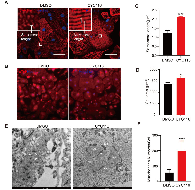
Fig. 2. CYC116 improves structural and mitochondrial maturation of H1-derived cardiomyocytes (H1-CMs).
(A and B) Representative images of immunofluorescence staining of α-Actinin and cTnt in DMSO- and CYC116-treated H1-CMs. Scale bars = 50 μm. (C and D) Sarcomere length (C) and cell area (D) measured from (A) and (B), respectively. Myofibrils with at least ten well recognized, continuous α-Actinin+ bands were measured and the length were divided by the number of sarcomeres. The cell area of cTnt+ H1-CMs is calculated by total fluorescence intensity/cell number (C) (n = 20-30 cells). (E) Transmission electron microscopy (TEM) images of H1-CMs treated with DMSO or CYC116. MF, myofibrils; Z, Z-bands; Zb, Z-bodies; Mit, mitochondria. Scale bars = 1 μm. (F) Quantification of mitochondria per CM with TEM images (n = 25 cells). Data are presented as the mean ± SEM. *P < 0.05; ****P < 0.0001.

