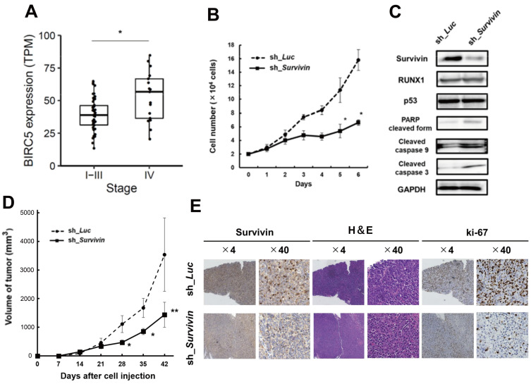
Fig. 2. Survivin knockdown inhibits the proliferation of MRT cells in vitro and in vivo.
(A) Boxplots of BIRC5 (survivin) in MRT stage I-III versus stage IV. The bottom and top of the box represent the first and third quartiles, respectively; the band inside the box represents the median. *P = 0.00663 by Mann–Whitney U test. TPM, transcripts per million. (B) Growth curves of MP-MRT-AN cells lentivirally-transduced with control (sh_Luc) or survivin shRNA (sh_Survivin) in the presence of 3 μM doxycycline (n = 3). (C) Immunoblotting of survivin, RUNX1, p53, apoptosis related proteins (poly(ADP ribose) polymerase [PARP], cleaved caspase 9, and cleaved caspase 3), and GAPDH in KP-MRT-YM cells with control (sh_Luc) or survivin shRNA (sh_Survivin). Cells were incubated with 3 mM of doxycycline for 48 h before being lysed for protein extraction. (D) Anti-tumor effects were examined by changes in the volume of xenograft tumor cells that knocked down survivin by shRNA (n = 6) or transduced control (n = 6). Data are represented as the mean ± SEM. *P < 0.05, **P < 0.01, by two-tailed Student’s t-test. (E) Representative microscopic images of tumor histology. Results obtained from staining and immunohistochemical staining with anti-human survivin antibody, H&E, and ki-67 are shown (original magnification ×4 and ×40 [insets]).

