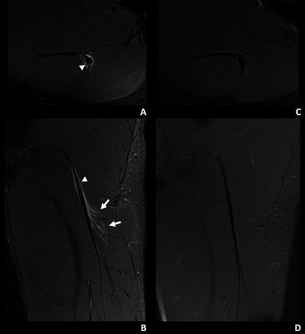
Fig. 1.

A 19-year-old left leg dominant football player of the non-reinjured group with mild tenderness months ago in the proximal portion of the left thigh. During a sprint in a match he experienced sudden pain in the proximal hamstrings region. A, B Axial and coronal T2-weighted fat-saturated images from a baseline MRI show a myotendinous injury of the proximal MTJ of the biceps femoris long head, I Pp 3r 0 (Muscle Location Grade and Reinjury [MLG-R] classification [33]). There is a mixed intratendinous tear (arrowhead) with peritendinous oedema and trace interstitial oedema (arrows). C, D Axial and coronal T2-weighted fat-saturated images from a control MRI, 48 days later, show a mature tendinous scar without oedema
