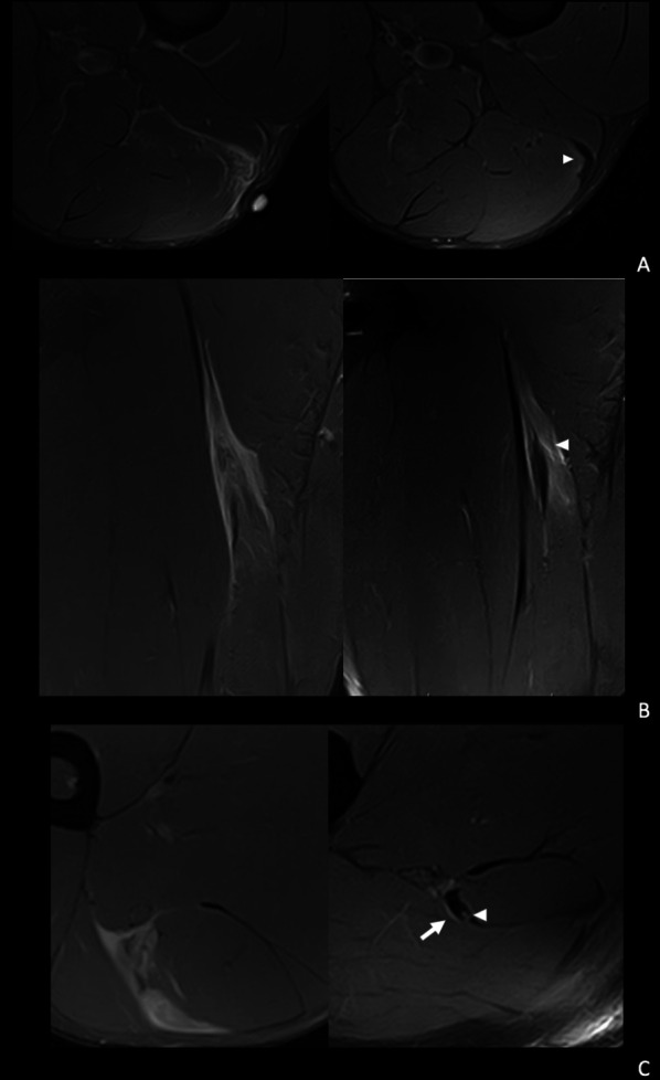
Fig. 4.

Spectrum of findings related to risk of reinjury. A A 31-year-old right leg dominant futsal player. Axial T2-weighted fat-saturated images shows: from a baseline MRI (left) a distal myotendinous injury of the left biceps femoris long head, I Dd 3r 0 (left). In the pre-RTP image (right) we can see a mature scar and a mild adaptive oedema (arrowhead). No signs associated with a higher reinjury risk. B 21-year-old right leg dominant football player. Coronal T2-weighted fat-saturated images from a baseline MRI show a proximal myotendinous injury of the left biceps femoris long head, I Pp 3r 0 (left). In the pre-RTP image (right) we can see a mature hypertrophic scar and interstitial oedema (arrowhead). This was considered medium risk for reinjury. C 22-year-old left leg dominant football player. Axial T2-weighted fat-saturated images from a baseline MRI (left) show a proximal myotendinous injury of the left biceps femoris long head, I Pp 3r 1. In the pre-RTP axial T2 image (right) we can see proximal to the previous injury tendinous tear (arrowhead) and intermuscular oedema (arrow). This was considered high risk for reinjury
