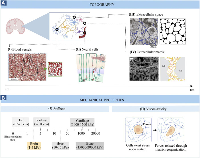
FIGURE 1.
Topography (A) and mechanical properties (B) of brain tissue. Images from panel A are adapted from (I) Microvascular networks in the human brain (Peyrounette et al, 2018), (II) Schematic representation of the developing cortical plate (Barry et al, 2014), (III) Electron micrograph of mouse cerebral cortex where the extracellular space is coloured in blue (left) ((Korogod et al, 2015) and computer generated configurations of the brain extracellular space (right) (Chen and Nicholson, 2000); (IV) Scanning electron micrograph of the brain ECM (left) (Tajerian et al, 2018) and schematic representation of the ECM (right) (Odackal et al, 2017). (B) represents the stiffness range (I) and viscoelasticity (II) of brain tissue.

