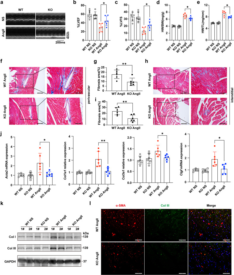
Fig. 3. KDM5B deficiency prevents pressure overload-induced cardiac dysfunction and cardiac fibrosis.
a Representative echocardiographic M-mode images of left ventricles from KDM5B-KO or littermate control WT mice on Day 28 after AngII or normal saline (NS) infusion. b, c Echocardiographic measurement of the LVEF (b) and LVFS (c) of KDM5B-KO or WT mice on Day 28 after AngII or NS infusion (n = 6 mice per group). d, e The ratio of heart weight to body weight (HW/BW) (d) and the ratio of heart weight to tibia length (HW/TL) (e) of KDM5B-KO or WT mice on Day 28 after AngII or NS infusion (n = 6 mice per group). f–i Representative Masson’s trichrome images and quantitation of perivascular (f, g) or interstitial (h, i) fibrosis in myocardial tissues from KDM5B-KO or WT mice on Day 28 after AngII or NS infusion (n = 6 mice per group). Scale bar, 200 μm (left), 100 μm (right). j Q-PCR analysis of Acta2, Col1a1, Col3a1 and Ctgf mRNA expression levels in myocardial tissues from KDM5B-KO or WT mice on Day 28 after AngII or NS infusion (n = 6 mice per group). k Immunoblot analysis of Col I and Col III protein expression in myocardial tissues from KDM5B-KO or WT mice on Day 28 after AngII or NS infusion. l Representative immunofluorescence staining of α-SMA (red) and Col III (green) in myocardial tissues from KDM5B-KO or WT mice on Day 28 after AngII infusion. Scale bar, 50 μm. *p < 0.05, **p < 0.01. Unpaired Student’s t test (g, i) or one-way ANOVA (b–e, j) was performed.

