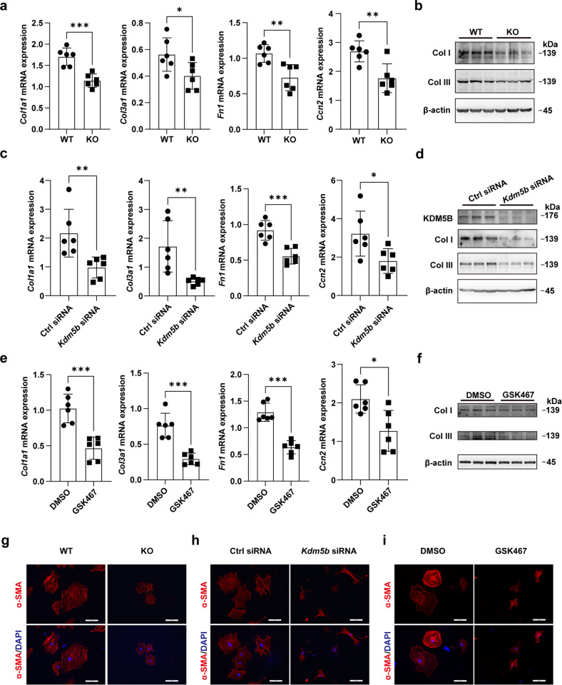
Fig. 4. KDM5B promotes fibrotic responses and the transition of cardiac fibroblasts.
a, b Q-PCR analysis of Col1a1, Col3a1, Fn1 and Ccn2 mRNA expression (a) (n = 6 per group) or immunoblot analysis of Col I and Col III protein expression (b) in KDM5B-deficient (KO) or littermate control WT cardiac fibroblasts stimulated with TGF-β (10 ng/ml) for 24 h. c, d Q-PCR analysis of Col1a1, Col3a1, Fn1 and Ccn2 mRNA expression (c) (n = 6 per group) or immunoblot analysis of Col I and Col III protein expression (d) in Kdm5b-silenced or control siRNA-transfected cardiac fibroblasts stimulated with TGF-β (10 ng/ml) for 24 h. e, f Q-PCR analysis of Col1a1, Col3a1, Fn1 and Ccn2 mRNA expression (e) (n = 6 per group) or immunoblot analysis of Col I and Col III protein expression (f) in cardiac fibroblasts treated with the KDM5B inhibitor GSK467 or DMSO followed by stimulation with TGF-β (10 ng/ml) for 24 h. g–i Representative immunofluorescence staining of α-SMA (red) in cardiac fibroblasts with KDM5B-deficiency (g), KDM5B knockdown (h) or GSK467 treatment (i) and the corresponding control cardiac fibroblasts (g–i) stimulated with TGF-β (10 ng/ml) for 24 h. Similar results were obtained from three independent experiments (g–i). Scale bar, 50 μm. *p < 0.05, **p < 0.01, ***p < 0.001. Unpaired Student’s t test (a, c, e) was performed.

