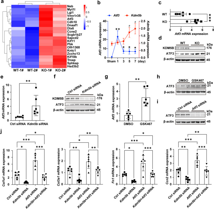
Fig. 7. KDM5B promotes cardiac fibrosis by inhibiting ATF3 expression.
a Heatmap showing the top 20 upregulated differentially expressed genes in myocardial tissues from KDM5B-KO and littermate control WT mice on Day 7 after MI surgery. b Q-PCR analysis of Atf3 and Kdm5b mRNA expression in the myocardial tissues of WT mice on the indicated day after MI or sham operation (n = 6 per group). #p < 0.05 vs. Kdm5b mRNA levels in the sham operation group; *p < 0.05, **p < 0.01 vs. Atf3 mRNA levels in the sham operation group. c, d Q-PCR analysis of Atf3 mRNA (c) (n = 6 mice per group) or immunoblot analysis of ATF3 protein levels (d) in myocardial tissues from KDM5B-KO or WT mice on Day 7 after MI surgery. e, f Q-PCR analysis of Atf3 mRNA expression (e) (n = 6 per group) or immunoblot analysis of ATF3 protein expression (f) in Kdm5b-silenced or control siRNA-transfected cardiac fibroblasts stimulated with TGF-β (10 ng/ml) for 24 h. g, h Q-PCR analysis of Atf3 mRNA expression (g) (n = 6 per group) or immunoblot analysis of ATF3 protein expression (h) in cardiac fibroblasts treated with the KDM5B inhibitor GSK467 or DMSO followed by stimulation with TGF-β (10 ng/ml) for 24 h. i Immunoblot analysis of ATF3 protein expression in cardiac fibroblasts transfected with Atf3 siRNA or control siRNA. j Q-PCR analysis of Col1a1, Col3a1, Fn1 and Ccn2 mRNA expression levels in cardiac fibroblasts transfected with Kdm5b siRNA, Atf3 siRNA, control siRNA or cotransfected with Kdm5b and Atf3 siRNA followed by stimulation with TGF-β (10 ng/ml) for 24 h (n = 6 per group). *p < 0.05, **p < 0.01, ***p < 0.001. Unpaired Student’s t test (c, e, g) and ANOVA (b, j) were performed.

