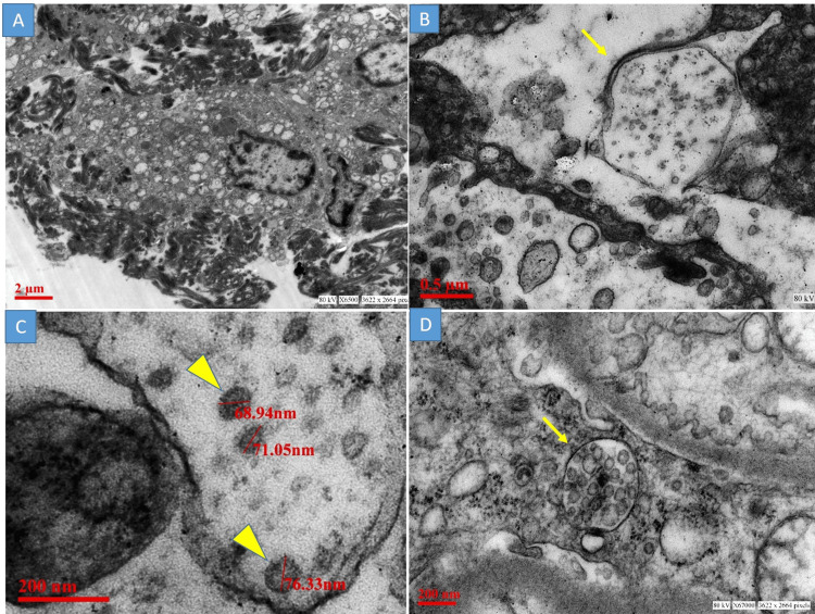
Figure 3. Transmission electron microscopy.
A: Type II pneumocyte with surface fibrin material; B: Viral particles were observed in double membrane-bound vesicles (yellow arrow), C: Aggregates of round enveloped viral particles of size 68nm to 80nm. Note a few electron-dense dots corresponding to transversely cut helical nucleocapsid (yellow arrowhead); D: A multivesicular body mimicking the viral particles (yellow arrow)

